
Visualization
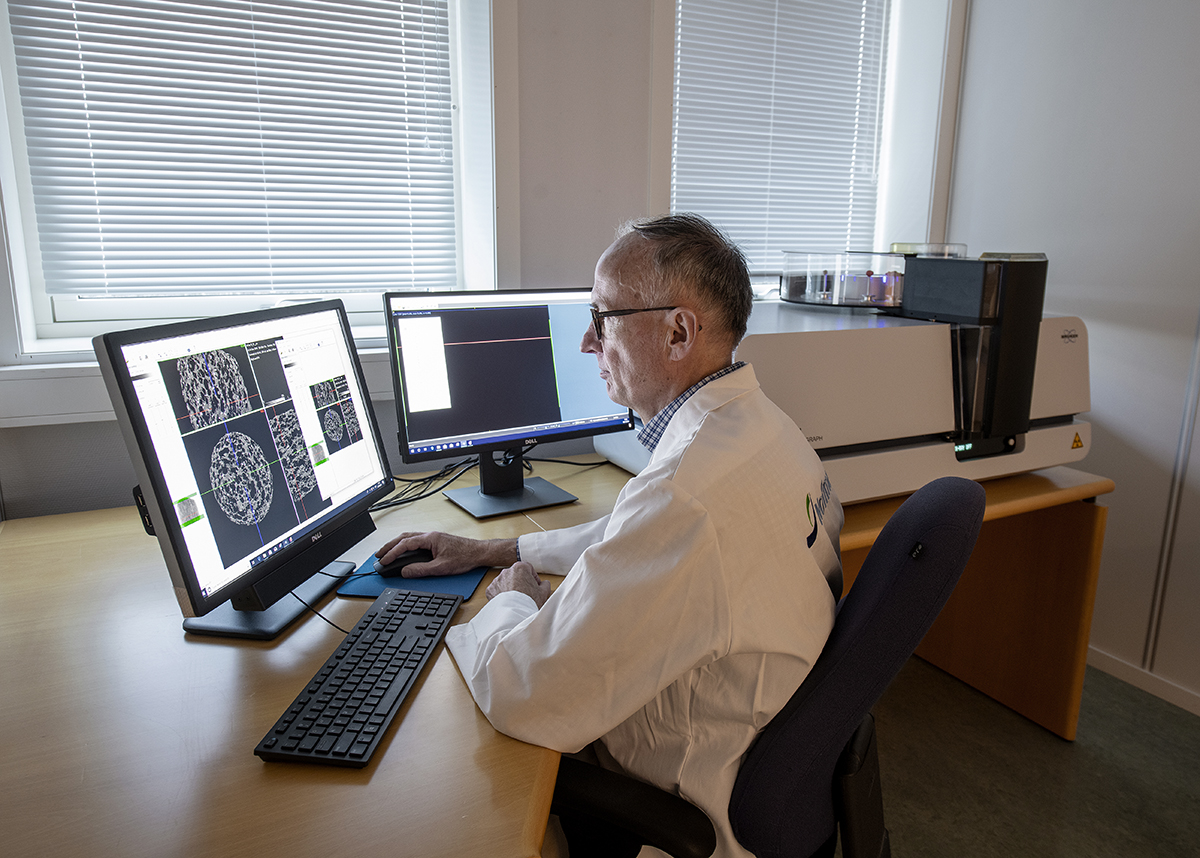

CT-scanner. Photo: Helge Skodvin @ Nofima.
The laboratory includes equipment for imaging of the macro, micro and internal 3-D structure of different objects.
Available equipment:
- X-ray Micro Computed Tomography (CT-scanner) enabling non-destructive imaging of the internal 3-D microstructure of an object. Equipped with an automatic sample changer. Maximum sample size up to 96mm in diameter and length 120mm. Resolution down to 5 µm. Cooling, heating and compression/tension stages are available.
- Stereo microscope with digital camera; magnification 6,3 – 80x.
- Digital SLR camera with close-up lens (1x).
